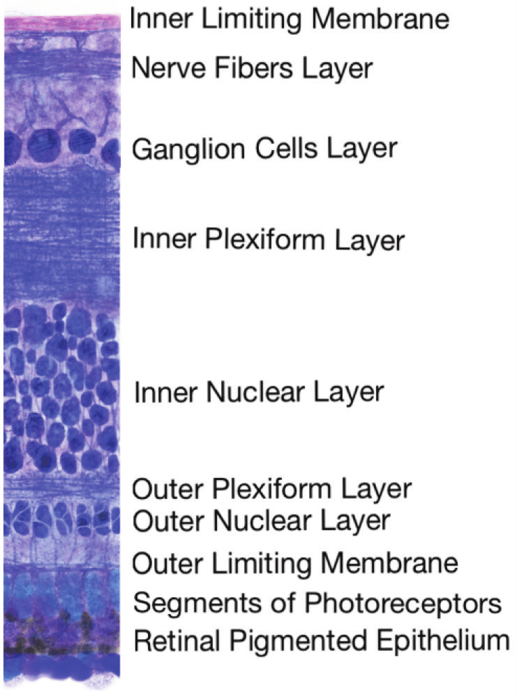
The retina is the thin layer that lines the back of the eye internally, it located near the optic nerve. The function of the retina is to receive the light that the lens has focused, convert it into neural signals, and send these neural signals to the brain via the optic nerve for visual recognition. The retina is the innermost layer of the wall of the human eye. It is in close contact with the vitreal cavity on one side and with the choroid (of the uveal layer) on the other side.
The layers of the retina (outer to inner) are as follows:
The Retinal Pigment Epithelium - is adjacent to the choroid, the pigment epithelium absorbs light to reduce back reflection of light onto the retina. The Photoreceptor layer - contains photosensitive outer segments of rods and cones. External (Outer) Limiting Membrane - maintains the structure of the retina through mechanical strength. The Outer Nuclear Layer - contains cell bodies of the rods and cones. The Outer Plexiform Layer - contains synapses between axons of photoreceptors and dendrites (projections of a nerve cell that receive signals from other nerve cells) of intermediate neurons. The Inner Nuclear Layer - contains cell bodies of intermediate neurons and muller cells. The Inner Plexiform layer - contains synapses between intermediate neurons and ganglion cells of the optic tract. The Ganglion Cell Layer contains cell bodies of ganglion cells. The Nerve Fiber Layer - consists of the axons of the ganglion neurons. The Inner Limiting Membrane - is the structural interface between the retina and the vitreous. It is the basal lamina (a layer of extracellular matrix secreted by the epithelial cells) of the inner retina which is formed by the footplates of muller cells.

Add comment
Comments